A pathologist examines a tissue section under a microscope. The tissue has been fixed in formalin, embedded in paraffin, sliced into 4-micron sections, mounted on a glass slide, deparaffinized, stained with hematoxylin and eosin, coverslipped, and dried. This process takes 12-48 hours, consumes the tissue section irreversibly, and produces a single stain. If the pathologist needs a different stain - PAS for kidney, trichrome for liver fibrosis, HER2 IHC for breast cancer - another section must be cut, another round of chemical processing completed, another day added to the turnaround.
Virtual staining asks a simple question: what if you could skip all of that?
The idea is to capture the intrinsic spectral signature of unstained tissue using hyperspectral or autofluorescence imaging, then use deep learning to generate images that are visually indistinguishable from chemically stained tissue. No chemicals. No tissue destruction. No waiting. And because the tissue is never consumed, you can generate unlimited virtual stains from the same physical section - H&E, IHC, special stains - all computationally, all from one acquisition.
This is not a speculative concept. In 2025, board-certified pathologists could not distinguish AI-generated virtual stains from real chemical stains in blinded evaluations. The technology is commercially available for research use, with clinical validation underway. The question is no longer whether virtual staining works - it is how fast the regulatory and clinical adoption pathways will move.
This article covers how hyperspectral imaging works in the pathology context, the deep learning architectures that power virtual staining, the clinical evidence across cancer types, and the software infrastructure required to deploy this at scale.
The Staining Problem
Histopathological staining has not fundamentally changed since Paul Ehrlich introduced aniline dyes in the 1870s. Hematoxylin binds to nucleic acids (staining nuclei blue-purple). Eosin binds to proteins (staining cytoplasm and stroma pink). The result is the ubiquitous H&E slide that pathologists have trained on for 150 years.
The problems with chemical staining are well known:
Time. Standard histology processing takes 12-48 hours from tissue receipt to stained slide. Rapid processing protocols exist (1-2 hours) but sacrifice quality. Intraoperative frozen sections take 20-30 minutes but introduce freezing artifacts.
Variability. Staining intensity and quality vary between labs, between technicians, between batches of reagent, and between days. Immunohistochemical staining (IHC) is even more variable - antibody lot-to-lot differences, antigen retrieval variability, and detection system sensitivity all contribute. This variability reduces diagnostic reproducibility and complicates AI training on multi-center datasets.
Tissue consumption. Each stain destroys one section of tissue. For core needle biopsies with limited material, the pathologist must choose which stains to order carefully. Once the tissue is consumed, restaining is impossible. This is particularly problematic in molecular diagnostics workflows where tissue must be preserved for genomic sequencing.
Cost. Routine H&E staining costs $5-15 per slide. IHC stains cost $50-200 per antibody per slide. A breast cancer workup requiring H&E plus ER, PR, HER2, and Ki-67 IHC runs $250-800 in staining costs alone, plus pathologist interpretation time. Multiply across millions of cases annually, and histology staining is a billion-dollar cost center.
How Hyperspectral Imaging Works in Pathology
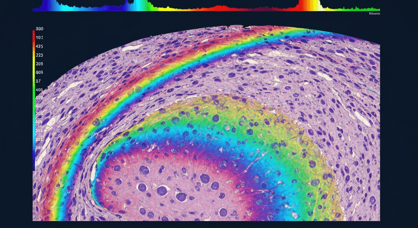
Standard microscopy captures three color channels (RGB) - a lossy reduction of the continuous visible spectrum into three broad bands. Hyperspectral imaging captures tens to hundreds of narrow spectral bands across the visible and near-infrared range, producing a data cube where every pixel has a full spectrum.
In tissue, this spectral richness encodes molecular information that RGB cannot distinguish. Hemoglobin, collagen, lipids, melanin, NADH, and flavins all have distinct spectral absorption and autofluorescence signatures. A hyperspectral image of unstained tissue contains enough molecular contrast to differentiate tissue types, identify cellular structures, and detect pathological changes - all without staining.
Acquisition Modes
Three optical configurations are used for hyperspectral pathology imaging:
Pushbroom (line-scan). A slit selects one spatial line. A dispersive element (prism or grating) spreads each point along the line into its spectral components. The detector captures a full x-lambda image in one frame. The sample stage translates to build up the spatial y-dimension. This produces the highest spectral resolution (150+ bands) and the most complete data, but requires mechanical scanning and is slower.
Snapshot (mosaic filter). A filter array on the detector splits each pixel into spectral sub-pixels, capturing all spatial and spectral information in a single sensor readout. IMEC's Snapscan technology achieves 150+ spectral bands at 3650 × 2048 pixels (7 megapixels) in the 470-900 nm range. A 2024 study found snapshot cameras are viable alternatives to pushbroom for real-time brain tissue identification. The trade-off is reduced spatial resolution per spectral band.
Tunable filter (LCTF or AOTF). A liquid crystal tunable filter or acousto-optic tunable filter selects one wavelength at a time, capturing the full field of view at each wavelength sequentially. Good spatial resolution at each wavelength, but slow due to sequential acquisition. A 2025 spatiotemporal filter array chip combines Fabry-Perot filters with liquid crystal modulation for simultaneous high spatial, temporal, and spectral resolution - a potential breakthrough in acquisition speed.
Spectral Range
Most pathology HSI systems operate in the visible to near-infrared range:
| Range | Wavelength | Key Chromophores | Tissue Penetration |
|---|---|---|---|
| Visible (VIS) | 400-780 nm | Hemoglobin, melanin, cytochromes | Surface (<1 mm) |
| Near-infrared (VNIR) | 780-1000 nm | Water, lipids, collagen | Moderate (1-3 mm) |
| Short-wave IR (SWIR) | 1000-2500 nm | Water, molecular bonds (NIR overtones) | Deep (3-10 mm) |
The 400-1000 nm range captures the strongest endogenous chromophore signatures and is where most clinical work is concentrated. Extending into the NIR adds molecular bond information - the same overtone and combination bands discussed in our NIR integration guide - at the cost of larger, more expensive detector arrays (InGaAs instead of silicon).
AI-Powered Virtual Staining
Virtual staining is a learned image-to-image translation task: given an input image of unstained or autofluorescence tissue, generate an output image that matches what the tissue would look like after chemical staining.
Architecture Evolution
The field has progressed through three generations of deep learning architectures:
Conditional GANs (2019-2023). Pix2Pix and its variants were the first architectures to produce convincing virtual stains. A generator network transforms the autofluorescence input into a virtual stain, while a discriminator network tries to distinguish real stains from virtual ones. The adversarial training produces sharp, realistic images. UCLA's Ozcan group demonstrated virtual HER2 IHC staining using conditional GANs - three board-certified breast pathologists found the virtual IHC images as accurate as real immunohistochemically stained counterparts.
CycleGAN (unpaired training). CycleGAN relaxes the requirement for pixel-aligned training pairs by enforcing cycle consistency - translating an image from domain A to B and back should recover the original. This is useful because perfectly aligned autofluorescence and stained image pairs are difficult to produce (the staining process itself changes the tissue). However, the strict cycle-consistency constraint can suppress nuanced morphological features, producing output that is smoother than real stains.
Diffusion models (2024-2025). Conditional diffusion models have overtaken GANs on PSNR, SSIM, and FID scores for virtual staining. StainDiffuser and related approaches generate virtual stains through iterative denoising, producing more stable and reproducible output than GANs. Ozcan's group published a pixel super-resolved virtual staining method using diffusion models in 2025, achieving a 4-5x increase in spatial resolution beyond the input image resolution - the virtual stain is actually higher resolution than the autofluorescence input.
Vision Transformers (2025-2026). ViT-Stain applies vision transformer architectures to virtual staining of skin histopathology, achieving 85% overall diagnostic concordance with virtual H&E stains and a Fleiss' kappa of 0.88 across pathologist evaluators. The global contextual learning of transformers captures long-range tissue architecture that CNNs miss.
Misalignment Problem
A fundamental challenge in training virtual staining models is spatial misalignment between the training pairs. The autofluorescence image is captured before staining. The H&E image is captured after staining, which physically deforms the tissue (dehydration, clearing, coverslipping). Even adjacent serial sections have structural differences.
A 2026 Nature Communications paper introduced cascaded registration mechanisms that separate image generation from spatial alignment. The result: expert pathologists cannot distinguish the virtual stains from real chemical stains. This was not possible with earlier architectures that required pixel-perfect alignment.
Clinical Evidence
Thyroid Cancer Detection
Thyroid is one of the most studied applications for hyperspectral pathology imaging, with multiple groups reporting consistent results:
- Infrared HSI + deep neural networks (2024): 93.66% accuracy, 93.47% sensitivity, 96.93% specificity via k-fold cross-validation for differentiating thyroid cancer from normal tissue.
- Adaptive spectral feature selection network (2025): Outperformed CNN, ViT, and SVM models for distinguishing Hashimoto's thyroiditis from papillary thyroid carcinoma, with the 400-500 nm wavelength range proving most discriminative.
- TimeSformer model on hyperspectral histological data (2024): Consistently outperformed RGB-based models for thyroid carcinoma margin assessment, demonstrating that the spectral dimension adds information that RGB color cannot capture.
- Multi-architecture comparison (2025): LDA model achieved 94% accuracy, 94% sensitivity, 95% specificity across thyroid tissue classification.
Brain Tumor Delineation
The HELICoID project used Headwall Hyperspec cameras (VNIR + NIR) for intraoperative brain tissue classification during neurosurgery. Deep learning pipelines achieved approximately 80% overall accuracy for multiclass classification (tumor core, infiltrating tumor, normal brain, blood). A two-layer pixel-wise DNN achieved 85% overall accuracy. The GLIMMER score - a decision-support metric derived from hyperspectral imaging - achieved AUC 0.95, sensitivity 94.7%, and specificity 83.3%, with 100% sensitivity in the glioblastoma subgroup.
For Raman-based intraoperative margin assessment (which uses spectral imaging principles but with a different modality), see our companion article on intraoperative Raman margin assessment.
Virtual Staining Concordance
The critical clinical metric for virtual staining is pathologist concordance - do pathologists reach the same diagnostic conclusions from virtual stains as from real chemical stains?
| Application | Architecture | Concordance | Source |
|---|---|---|---|
| Virtual H&E (general) | Diffusion model | Complete (pathologist blinded) | Ozcan/UCLA, 2025 |
| Virtual HER2 IHC (breast) | Conditional GAN | Equivalent HER2 scoring | BME Frontiers, 2023 |
| Lung transplant biopsy | Virtual staining network | 82.4% concordance | BME Frontiers, 2025 |
| Heart transplant biopsy | Virtual staining network | 91.7% concordance | BME Frontiers, 2025 |
| NASH scoring | HSI + deep learning | Steatosis 0.91, ballooning 0.84, fibrosis 0.65 | Modern Pathology, 2024 |
| Skin histopathology | ViT-Stain | 85% diagnostic concordance, kappa 0.88 | PMC, 2026 |
| Tissue regions (general) | Misalignment-resistant | Indistinguishable from chemical stain | Nature Comms, 2026 |
Concordance rates of 82-92% for complex diagnostic tasks and "indistinguishable" ratings in blinded evaluations represent clinically meaningful performance. For context, inter-pathologist concordance for H&E interpretation varies from 60-90% depending on the diagnostic category - virtual staining is approaching the range of human variability.
Immuno-Oncology Biomarkers
Verily (Google) published work in January 2025 combining autofluorescence hyperspectral microscopy with machine learning to generate virtual H&E plus a multiplex immunofluorescence panel (DAPI, PanCK, PD-L1, CD3, CD8) from unstained non-small cell lung cancer tissue. The system reproduced key morphologic features and biomarker expressions at both tissue and cell levels, enabling identification of critical immune phenotypes: tumor area, T-cell density, and PD-L1 tumor proportion score. If validated clinically, this could replace the multi-day, multi-stain IHC workflow for immunotherapy companion diagnostics with a single autofluorescence acquisition.
Companies and Products
Hyperspectral Hardware
| Company | Product | Configuration | Spectral Range | Resolution | Clinical Use |
|---|---|---|---|---|---|
| CytoViva | Enhanced darkfield HSI microscope | Pushbroom | VIS-NIR | High | Research - endogenous spectral analysis without staining |
| Headwall Photonics | Hyperspec VNIR A-Series | Pushbroom | VIS + NIR | 150+ bands | Research - used in HELICoID brain surgery project |
| IMEC | Snapscan | Snapshot + scanning | 470-900 nm | 150+ bands, 7 Mpx | Research - chip-level integration, blood smear imaging |
| Resonon | Pika L, Pika XC2 | Pushbroom | 400-1000 nm | 281 bands | Research - liver tissue ablation quantification |
| Photon etc. | IMA Hyperspectral | Global imaging | 400-1620 nm | VIS/NIR/SWIR | Research - faster than raster scanning systems |
Virtual Staining Software
Pictor Labs is the commercial leader in virtual staining. Co-founded by Aydogan Ozcan as a UCLA spinoff, the company raised $30M in September 2024 (led by Insight Partners and M Ventures). Their product portfolio:
- DeepStain / ReStain - high-fidelity virtual staining from a single tissue sample. Generates unlimited virtual stains from one acquisition.
- ClearStain (launched November 2025) - generates virtual H&E from unstained slides for molecular diagnostics workflows. In blinded evaluation, pathologists found virtually stained tissue visually comparable in 99-100% of regions reviewed. The key value proposition: preserves tissue for downstream molecular assays (genomic sequencing, FISH) while still providing morphological assessment.
All Pictor Labs products are currently For Research Use Only - no FDA clearance or CE mark.
Digital Pathology Platform Integration
Pictor Labs partnered with Proscia (Concentriq platform) in September 2025 to integrate virtual staining into the Concentriq digital pathology workflow. This is the first commercial pairing of virtual staining AI with a mainstream digital pathology platform.
Other digital pathology companies with AI capabilities (PathAI, Indica Labs/HALO) have not yet announced hyperspectral or virtual staining integrations, but the convergence is inevitable - PathAI expanded its AI diagnostic capabilities with a digital pathology analytics acquisition in June 2025, and Indica Labs received investment from Leica Biosystems in January 2025.
Software and Data Infrastructure
The Data Problem
A single hyperspectral image is massive. At 3600 x 2048 pixels x 150 spectral bands x 32-bit float precision, one frame is approximately 4 GB. A whole-slide hyperspectral image - covering the same area that a standard whole-slide scanner captures - can exceed several GB per slide. By comparison, a standard RGB whole-slide image is 1-3 GB.
Multiply by the number of slides per case, cases per day, and retention period, and the storage requirements are staggering:
Storage estimate for a mid-volume pathology lab:
  100 cases/day × 3 slides/case × 5 GB/slide = 1.5 TB/day
  365 days × 1.5 TB/day = 547 TB/year
  10-year retention = 5.5 PB

For comparison, the same lab's RGB whole-slide images:
  100 cases/day × 3 slides/case × 2 GB/slide = 600 GB/day
  365 days × 600 GB/day = 219 TB/year
  10-year retention = 2.2 PB
Compression is critical. Wavelet-based compression methods developed specifically for VNIR and SWIR hyperspectral data (2025) can achieve 10-20x compression ratios without diagnostically significant quality loss - but these are not standardized or widely adopted.
GPU Processing Requirements
Real-time virtual staining inference requires significant GPU resources:
- K-nearest neighbor classification on hyperspectral data: 30-66x speedup on GPU vs CPU
- K-means clustering: 150x speedup on GPU
- Full pipeline with PCA on multi-GPU: 180x speedup over serial processing
- Diffusion model inference for virtual staining: requires NVIDIA A100/H100 class GPUs for sub-minute generation
For a clinical deployment, the processing pipeline is:
Hyperspectral Acquisition (seconds to minutes)
    │
    ▼
Data Cube Preprocessing
    │  (spectral calibration, spatial registration, normalization)
    ▼
Virtual Staining Inference (GPU)
    │  (diffusion model or GAN, ~10-60 seconds per field of view)
    ▼
Image Tiling & Pyramid Generation
    │  (for whole-slide viewer compatibility)
    ▼
PACS / Digital Pathology Viewer
    │  (Proscia Concentriq, PathAI AISight, HALO)
    ▼
Pathologist Review
DICOM and Format Standards
There is currently no DICOM standard for hyperspectral pathology data. This is a significant gap. DICOM Supplement 145 supports tiled whole-slide images as multi-frame images at varying resolutions, but it was designed for RGB or single-channel images, not hyperspectral data cubes.
Hyperspectral data is typically stored in proprietary formats - ENVI, HDF5, or custom binary formats from each camera manufacturer. The virtual staining output (a synthetic RGB H&E image) can be stored as a standard whole-slide image in DICOM, TIFF, or SVS format. But the raw hyperspectral data - which is needed for reprocessing, restaining with different virtual stains, and quality assurance - has no standardized storage format.
As of 2024, the Leica Aperio GT 450 DX is the only whole-slide imaging system with FDA clearance for native DICOM output. Philips has announced an upcoming DICOM-native scanner but it is not yet cleared. For hyperspectral pathology, DICOM integration requires a custom DICOM wrapper around the vendor's native format.
Regulatory Status and Maturity Assessment
| Component | Maturity | Timeline Estimate |
|---|---|---|
| Hyperspectral imaging hardware | Commercial - research-grade instruments available from multiple vendors | Available now |
| Virtual H&E staining AI | Late research / early clinical validation - pathologist concordance studies complete | 2-3 years to regulatory submission |
| Virtual IHC staining AI | Research stage - proof of concept demonstrated, clinical validation needed | 3-5 years |
| End-to-end clinical platforms | Early clinical validation - Pictor Labs + Proscia integration available for research | 2-4 years to clinical use |
| FDA/CE cleared virtual staining | None - no submissions announced | Earliest 2028-2029 |
| DICOM/informatics integration | Nascent - no standards for HSI pathology data | Standards development needed |
There are currently no FDA-cleared or CE-marked hyperspectral pathology devices or virtual staining systems. Pictor Labs products are explicitly labeled For Research Use Only. Of the 51 FDA-authorized pathology AI devices through April 2026, only 7 are whole-slide imaging algorithms, and none involve hyperspectral imaging.
The market context is favorable. The global digital pathology market is projected to grow from $1.46B (2025) to $2.75B (2030) at 13.5% CAGR. Medical hyperspectral imaging diagnostics is the fastest-growing segment at 12.0% CAGR (2026-2033). The worldwide pathologist shortage - fewer trainees entering the field while case volumes increase - is a powerful adoption driver.
What This Means for Spectroscopy Software
Hyperspectral pathology is spectroscopy applied to tissue imaging at microscopic scale. The data pipeline challenges - spectral calibration, preprocessing, classification, result delivery - are the same ones we solve for point-measurement spectroscopy (FTIR, Raman, NIR), just at much higher data volumes.
The software requirements map directly:
- Spectral preprocessing → same algorithms (normalization, baseline correction, noise reduction), applied per-pixel across millions of pixels instead of per-spectrum
- ML classification → same model architectures (CNNs, transformers), applied to 2D+spectral data cubes instead of 1D spectra
- Result delivery → DICOM and PACS integration instead of HL7/FHIR, but the same pattern of delivering a classification result to a clinician
- Regulatory compliance → same IEC 62304, ISO 14971, and SaMD classification framework described in our IEC 62304 guide and SaMD classification article
The convergence of spectroscopy and digital pathology is not a future possibility - it is happening now. The teams that build the software infrastructure to handle hyperspectral data at pathology scale will define the next generation of diagnostic AI. The clinical workflow platform that connects spectral instruments to hospital systems is the foundation this infrastructure requires.
Further Reading
- Intraoperative Raman Margin Assessment - Raman spectroscopy in the operating room
- Building AI Pipelines for Spectral Classification - ML pipeline architecture for spectral data
- Building Clinical Workflow Software for Spectroscopy-Based Diagnostics - system architecture
- IEC 62304 for Spectroscopy Software - medical device software compliance
- Transfer Learning for Spectral Models - cross-instrument model adaptation
SpectraDx is clinical workflow software for spectroscopy-based diagnostics. We handle the integration layer between your spectrometer and your clinician. Learn more or get in touch.